Following is a paper presented for publication in a scientific journal. An introductory explanation is offered because of the unusually extensive scope that consists of a mechano-geometrical model of morphogenesis.
The paper provides the needed model for the science of biology much as did Dalton, Mendeleev, and Niels Bohr for chemistry. The reader is invited to partake in the further investigation of the model. The model may contradict work by developmental biologists who must either refute this solution to the development problem or accept it as scientific fact. "To disregard non-corroborative data is to falsify data."
____________________________________
THE FRACTAL GEOMETRY OF THE EMBRYO
Life is the crystallography of the lipid molecule.
The paper presented below is a mechanical model of embryogenesis, that is, an account of how the embryo is formed. The premise is based on the discovery of a geometrical algorithm that describes the path from one cell to complex embryo in the self-organizing patterns that occur in the mass of dividing cells, and how millions of species are encoded in the inequalities in the divisions of the egg.
This model of embryogenesis claims that the origin and diversity of living structural design is encoded in the inequalities of the first eight cells caused by inequalities in the first three cell divisions.
This paper identifies the physical phenomena responsible for the organization of the embryo and the diversity of the forms produced upon development. We demonstrate, that,
1. The embryo is a geometrical spiral fractal.
2. The embryo structure is encoded in the inequality of the first three cleavages of the egg causing physical inequalities in the first eight cells comprising the initial cubical matrix.
3. Embryogenesis is an exercise in fluid dynamics directed by the hydraulic interconnections among the first eight cells, and in the arcane frictionless and gravity-less laws of surface tension, whereby the lesser of the unequal pairs of daughter cells are subsumed by the greater.
Hence,
This model of embryogenesis claims that the origin and diversity of living structural design is encoded in the inequalities of the eight cells of the initial cubical matrix resulting from inequalities in the first three cell divisions.
___________________________________________________________________________
The Geometrical Origin of the Embryo in the Cubical Fractal Matrix F
Formed by the First Three Divisions of the Egg Cell
S. Pivar, P. Sheesley
PREFACE
In 1866 and 1874 Ernst Haeckel published his famous drawings showing that the embryos of all vertebrates from fish to man were quasi-identical. Ever since then scientists have never been able to explain this phenomenon or provide a coherent account to the interested legislative bodies when asked what an embryo is. This essay purports to shed light on the problem by describing the origin of the embryo both as a biological organ and a geometrical form. This paper demonstrates how biological structure is encoded in the embryonic cleavage pattern.
We claim:
The self-organized patterns assumed by the biological body originate in the hydrodynamic equilibrium patterns among the eight cells of the cubical matrix resulting from unequal cell division in the first three embryonic cell divisions. Taxonomic determination is encoded in the degree of inequality of the unequal divisions.
We suggest that:
The advent of inequality in the third division produces three-dimensional growth of cancer cells.
In the subject theorem the genes notably play no role. As Stephen Jay Gould stated in “Ontogeny and Phylogeny,” 1976: “The genes provide protein in precisely timed, measured doses, thus preserving and occasionally changing the otherwise immutable proportions of the phyletic body.”
ABSTRACT
Science has always presumed that the assembly of the body is guided by a priori knowledge of the outcome encrypted in some kind of code. The 1951 discovery of the wondrously complex structure of the DNA molecule fit the bill and set out the race to decode it. Yet twenty-five years later Richard Lewontin demanded, “Where is the gene for my nose?”. Charles Darwin’s ingenious suggestions failed to prove themselves. Now the new discipline of mechano-biology claims the mechanically- directed self-organization of the complex animal body.
This is the presentation of the discovered mechano-geometrical algorithm demonstrating that:
The self-organized patterns assumed by the biological body originate in the hydrodynamic equilibrium patterns among the eight cells of the cubical matrix resulting from the first three embryonic cell divisions, of which two are unequal. Taxonomic determination is encoded in the degree of inequality of the unequal divisions.
The prime mover is in the law revealed by interconnected balloons in the famous physics demonstration where principles of fluid dynamics are demonstrated by showing counterintuitive responses to forces. The origin of biological form is solved by the” eight-balloon experiment.”
Since the eight-cell cube results from inequalities in the first cleavages, the cells are composed of four pairs of unequal size.
We demonstrate as a corollary that the form of the embryo is that of a Mandelbrot Set Spiral Fractal, the reason for which is that the embryonic cells quantify expansion by the subdivision in rounds of the three axes of space, a necessary and sufficient condition for the generation of the geometrical fractal figure. Hence, “embryo” is a name of a figure in space comparable to triangle, square, sphere, pyramid, cube, or dodecahedron.
The mechanical laws governing cell cleavage cause sequential ninety-degree rotation of the cleavage axis causing the quantifying of the growth border, providing sufficient condition for fractalian expansion. Indeed, we are fractals. We grow in rounds of three, each ninety degrees from the previous.
This hypothetical model solution to the problem of how the embryo is made is presented with proof by Euclidian congruence with observed nature. The parallel is drawn with the quest for the atomic model solved in 1923 by the then seemingly preposterous model of Niels Bohr.
Another corollary to this paper is in its value as a fate map trajectory from ovum to embryo to oncologists who investigate the premise that cancer can be caused by mechanical disruptions of the early embryo-geometrical patterns. We propose unequal cleavage as a cause of cancer.
KEY TERMS
Oocyte, germ cell, soma, cleavage, mechanobiology, hydrodynamic, gastrulation, totipotent, surface tension fractal, Mandelbrot Set, fluid mechanics.
INTRODUCTION
While at the human scale Newtonian mechanics is ruled by gravity and friction, cells are moved by surface tension and electromagnetic forces, producing many unfamiliar phenomena. These underlying causative phenomena are described in this model of the life cycle.
Life became possible with the advent of the highly polar non-binary lipid molecule, capable of intercalary growth, and the formation of bilayer spheres that alternately grow and split in two sequentially through the three axes of space.
It is the mechanical and geometrical exigencies that derive from this bio-physical given that generate the forms of nature.
Life is the crystallography of the lipid molecule.
ARGUMENT
The first three cleavages of the egg create the eight-cell cubical matrix, basis of the left-right, head-tail, back-belly structural basis of bilateral animal life.
The Cubical Fractal. Cell division by Sachs’s Rule is the quantifying of cell division by rounds of divisions through the three axes of space. The first round of three divisions creates a cube. The second round creates a cube of cubes, and so forth ad infinitum, in the establishment of a Mandelbrot-series cubical/spiral fractal pattern, the basis of the morphology of bilateral animal life. Hence, the embryo is a fractal, the inevitable topological trajectory of an expanding spherical bilayer, in the periodical, sequential inversion of the surface preserved eternally in the perpetually regenerating germ membrane.
The fractal figure of which complex life is based is generated by the sequential rounds of cleavages through the three axes of space that generate a cubical octet of cells. The periodical repetition of the rounds of three cleavages creates the cubical fractal, consisting of cubes made of cubes made of cubes.
Unequal cleavages cause the spiral fractal symmetry of the bilateral animal body-forms.
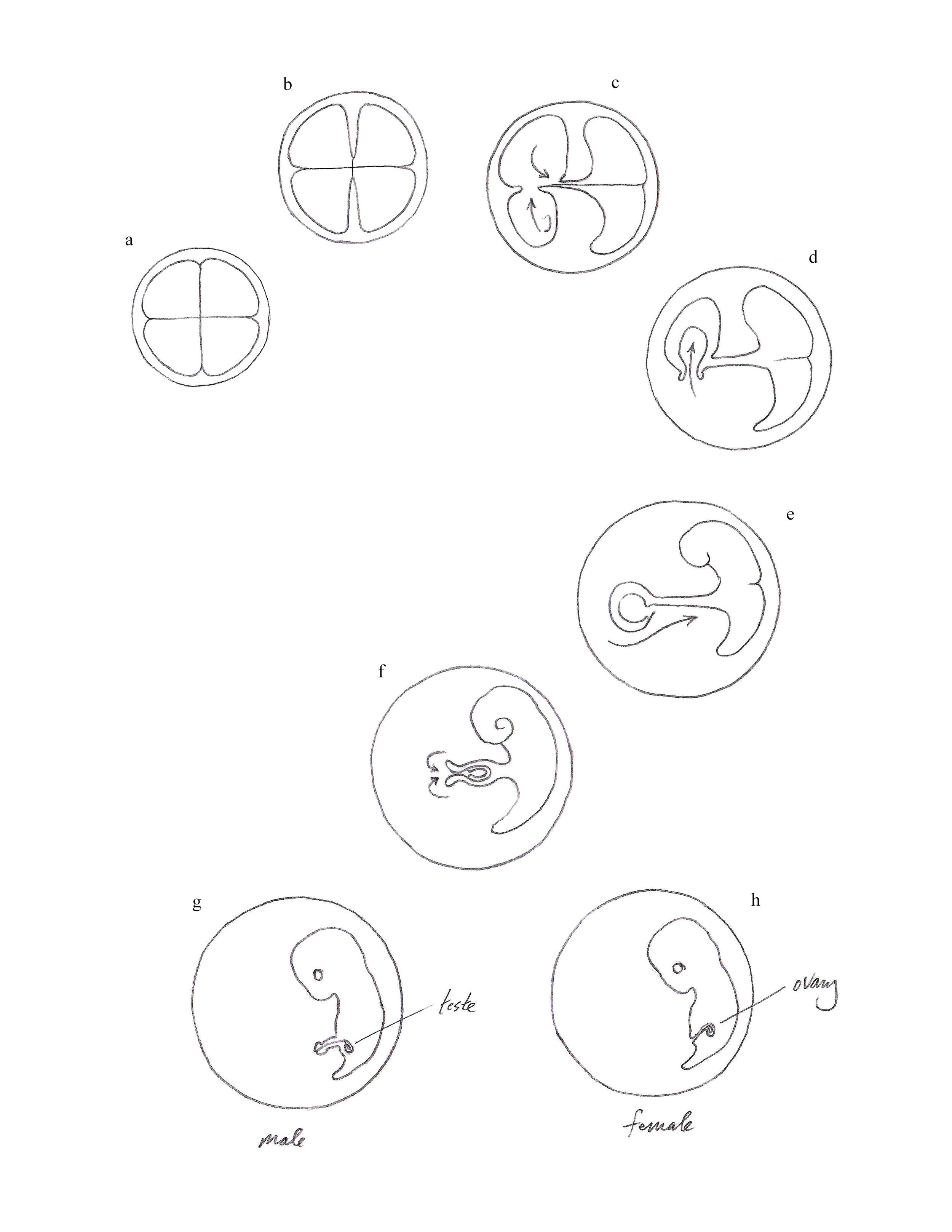
Morphogenesis. In three consecutive cleavages the egg cell is subdivided in four left-right pairs. The anterior pair of quadrants are the ancestors of the immortal germline body. The posterior quadrants are the embryonic ancestor of the mortal somatic body; the dorsal is the head, the ventral is the body. Of the first two oocytes, the larger is the progenitor of the germline and the smaller the progenitor and shaper of the somatic (i.e., mortal) body. The germ body periodically divides its eternal pattern, regenerating its lost half. The soma membrane shrinks, forming the embryo.
Gametogenesis is the invasion of the genetic hemisphere into the embryo in a sequence of steps driven by the hydraulic dynamic forces.
a. The anterior-ventral pair shrink to become testes and ovum within an embryonic sac and are drawn into the anterior-dorsal pair.
b. The anterior-dorsal pair are drawn into the embryo as the ovum.
c. In the male the interconnecting tube forms the male organ.
d. In the female the interconnecting tube is pulled inside-out, forming the female organ.
Hydromechanical Equilibrium
The first cleavage is equal.
The following two cleavages are unequal.
Unequal cleavage of two of the three axial cleavages generates the spiral fractal pattern of the embryo.
Incomplete cleavage preserves a capillary microtubule interconnection between the divided cells that can transport fluid and regulate pressure.
The incomplete and unequal cleavage of a growing bilayer sphere in the rounds of three cleavages in the three axes of space generates the structure of a cubical matrix of mutually hydraulically interconnected cells about a central plenum.
Hydraulic equilibrium is established in the cubical matrix as lesser cells of unequal pairs empty into the greater.
The establishment of Hydromechanical Equilibrium among the eight progenitor oocyte cells by intercellular fluid exchange produces morphological dissimilarity among the eight.
Spherical Transcription
The growth of the plenum generates the blastosphere, or blastocyst. The expansion of the blastocyst causes the transcription of the cuboid structure into the two-dimensional patterned pavement of the blastocyst. The blastocyst is paved with the spiral fractal growth pattern reduced to two dimensions. The proliferating octets of cells pave the periphery of the growing central plenum, called the blastocyst, whereby the blastula sphere becomes the spherical transcription of the cubical fractal structure.
Segmentation of the blastula results from tension caused by the growth of the blastocyst.
The claim is made here that the egg cell, that as an expanding bilayer, upon fertilization continues an eternal path of alternation of forms, as the sequential inversion of curvature. Observed comparative embryology offers myriad examples of development consisting of alternating inversions of the body morphology, most evident in the invertebrates. Noted are the larval stages of many orders of insects passing through stages of egg, grub, caterpillar, imago, chrysalis, adult. Periodic curvature inversion may be observed widely to account for the extravagant morphologies in marine larvae. It is noted that the inversion cycle is abbreviated in vertebrates by the sealing of the nerve cord.
The theorem is based on the mechano-geometrical axiom that upon expansion a curved bilayer surface tends to reverse its curvature due to the difference in the tension of the layers. Hence, an expanding spheroidal bilayer surface predictably proceeds to turn inside-out sequentially.
This theorem solves the conundrum posed by Ernst Haeckel in the famous drawings of the adult vertebrate families from fish to man, all deriving from an indistinguishably identical embryo.
In the above theorem the genes notably play no role. As Stephen Jay Gould stated in “Ontogeny and Phylogeny,” 1976: “The genes provide protein in precisely timed, measured doses, thus preserving and occasionally changing the otherwise immutable proportions of the phyletic body.”
CONCLUSION
The transparency of embryonic tissue continues to obscure the process called gastrulation which forms the embryo from centuries of inquiry of developmental biologists. This paper purports to offer a model solution to the problem of how the embryo is made and how it is preserved intact over eons.
The Lifecode: The downstream differentiation of plants and animals into millions of species is encoded in slight inequalities in the first three cleavages of the egg.]
REFERENCES
Gould, S.J. Ontogeny and Phylogeny. Belknap Press of Harvard University Press, 2003.
Lewontin, R.C. It Ain’t Necessarily So: The Dream of the Human Genome and Other Illusions, New York Review of Books, New York, 2000.
McCoy, R.C., Summers, M.C., McCollin, A. et al. Meiotic and mitotic aneuploidies drive arrest of in vitro fertilized human preimplantation embryos. Genome Med 15, 77 (2023). https://doi.org/10.1186/s13073-023-01231-1.
Parajón, Eleana, Alexandra Surcel, and Douglas N. Robinson. The mechanobiome: a goldmine for cancer therapeutics. American Journal of Physiology–Cell Physiology, Vol. 320, No. 3, 09 MAR 2021, https://doi.org/10.1152/ajpcell.00409.2020.
https://theconsciousvibe.com/are-humans-fractals-biology-and-behavior-the-science/.
ILLUSTRATIONS
The following small number of illustrations are drawn from the
Embryo Geometry Library of Illustrations accessible at
wwww.embryogeometry.com

Cuboid Fractal-Genesis
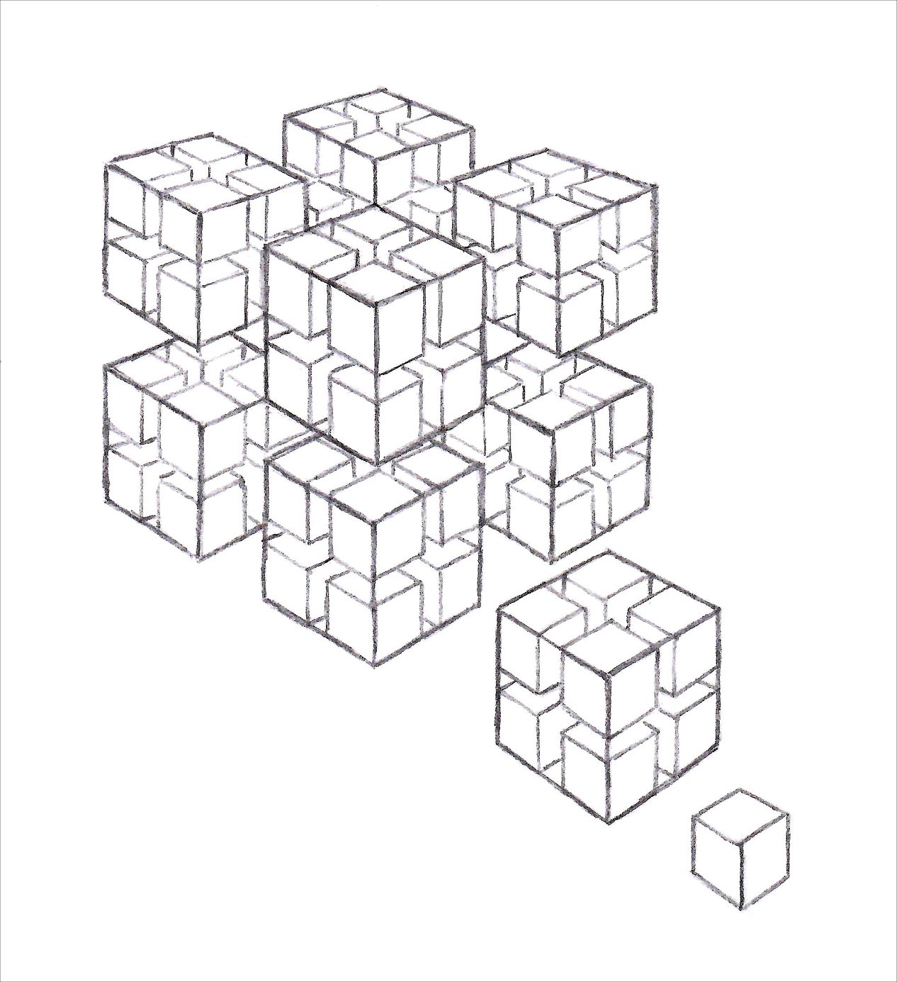
Cuboid Fractal-Genesis

Curvature Inversion and Embryogenesis

Embryogenesis and Sex Determination
Hydrodynamic Formation of the Somatic and Genetic Bodies

Ovum, Cleavage, Inversion
_____________________________________________________________________

RHINOCEROS V. RHINOCEROS
The Search for the Urform
What is the mutual characteristic that permits Mickey Mouse, Donald Duck, Bugs Bunny, Jiminy Cricket and SpongeBob SquarePants to deal with each other in universal anthropomorphism? Answer: Despite extreme diversity in animal bodyform, all animals act the same way.
Behavior is mature at birth and universally uniform in all animal species. Behavior experiences no development. Except for humans and a few other primates, there is no learning curve in animals. Flies are born aces at flying and do not improve with practice. That which evolutionary biologists call LUCA (the Last Universal Common Ancestor) must be behavior. Everything else is diverse.
The LUCA, then, must be the behavior of the last common morphological structure, which is, of course, the blastula, that primal ball of cells resulting from the serial geometrical bisection of the egg cell—which gives way to the embryo in the embryological process called gastrulation.
The blastula turns into the embryo but seems to keep the uniform blastula behavior of all animals. Behavior must originate in the blastula and be conserved in the embryo. Although all embryos are different, all blastulae are of the same predictable, geometrically regular, construction.
The blastula is composed of concentric spherical gridded membranes that periodically reproduce themselves, sloughing off the outer layers in adults in different ways–from the skins of snakes, insect imagoes, and in the human bath.
Hence, morphological diversity and behavioral uniformity are the chief features of the Animal Kingdom.
EMBRYO GEOMETRY
the science of understanding the patterns we see in dividing cells
The origin of the shapes of plants and animals lies in the patterns that form geometrically in dividing embryonic cells. The form of the human body derives from natural self-organization much as does the rainbow, the wave and the daisy. Genus and species are the result of the different ways cells can form a ball.
How does nature shape plants and animals? How does evolution work? The recent history of the science of evolution and embryology has reviewed and rejected natural selection and the DNA code as candidates. Today the sole theory of interest is mechanobiology, pursued by thousands of investigators in hundreds of organizations. A common belief is that the embryo is formed by the self-organization of patterns in the dividing cells. Yet the failure to make head nor tail of the first embryonic structures has defied the direct observation of the process. Embryogenesis remains a mystery.
The model proposed is the result of over thirty years of investigation with the participation of numbers of scientists and illustrators. It is presented as “blueprints” for the making of virtually all plant and animal body and surface forms in over a thousand mechanical drawings and photographs.
The path from one cell to adult human is depicted in simple animations where each step is caused mechanically by the conditions of the previous step. The intuitively comprehensible mechanical and geometrical processes are presented without mathematical equations or chemical formulae.

Embryo Geometry, The Model
The vertebrate embryo is the shriveled remnant that results from the bursting of the simple swollen, balloon-like blastula, much as the prune is a dried plum. (The embryo of a fish cannot be readily distinguished from that of the human.)
The following is a hypothetical model based on the predictable, plausible, mechanical consequences of the bursting of the swollen bilayer blastula.
The hypothesis we present is that the embryo is formed by the elastic recoil of the balloon-form blastula membrane upon bursting failure. Spherical, cylindrical and ovoidal blastulae produce respectively the radial, vermiform and bilateral body forms. Small mechanical variants in the action are the cause of taxonomic variation.
THE STEPS OF EMBRYOGENESIS
1. The swollen spheroidal bilayer blastula bursts by splitting from pole to pole along the future ventral midline;
2. The tense blastula membrane recoils dorsally;
3. The entire dorsal half of the blastula membrane is forced into the dorsal midline that seals over, forming the nerve cord and the central nervous system. The neurons, newly formed by the compression of the blastocysts, extrude into the brain case of the forming skull;
4. The model hypothesizes that the blastula membrane is subdivided in a number of self-organized circumferential banded girdles. This supposition provides a model for the shaping of the vertebrate torso, limbs, and skull, as well as the segmented insect and crustacean body;
5. The development of the outer layer of the blastula accounts for a multitude of structures including human hair patterns, the shell of the turtle and armadillo, and the many bizarre dorsal structures on the dinosaurs.

The outer layer of the blasula forms a variety of dorsal structures in the dinosaur.

A detailed illustration showing recoil from the ventral midline, forming the neural tube on the dorsal side. The dorsal tube extrudes into the brain case forming the brain and sense organs as the skull segments form the structure of the skull.

The axioms of self-organizations include: 1) initial subdivision in three axes; 2) the basic division into a hollow animal blastula or a solid plant sphere; 3) plant growth through multilayer telescoping; 4) animal growth in either radial or bilateral models; 5) the principles of multilayer growth; 6) superficial patterning as a result of geometrical self-organization.

An overview showing the extrusion of the neural tube into the brain case and its expansion, forming the brain.
A Summary of the Theory of Embryo Geometry
The heart of biological science is the search for the way the embryo is formed. The rest is known.
This proposed model is an account of embryogenesis, tantamount to the blueprint for the assembly of the embryo.
Included are the phenomena of morphogenesis, organogenesis, segmentation, limb development, and nerve cord development, all the demonstrable consequence of the singular event of gastrulation. The model is illustrated by hundreds of mechanical animation drawings.
The model describes the instantaneous catastrophic embryological event called gastrulation as the bursting of the first embryonic structure, the blastula, and its elastic recoil as demonstrably the universal origin of animal form.
The embryo is the deflated form of the tense, balloon-like blastula membrane upon bursting and elastic recoil.
Organogenesis is the deformation by elastic recoil of the pattern of self-organized circumferential girdles that encompass the blastula from pole to pole.
The radial, vermiform, and bilateral body forms result, respectively, from a spherical, cylindrical, or ovoidal blastula. Morphogenesis is the anterior-dorsally directed migration upon bursting failure of the separate layers of the membrane bilayer, governed by the mechanical exigencies of surface wrinkling pattens. Embryogenesis is predictable by the mathematics of topological surface wrinkling patterns.
Taxonomic differentiation is the result of mechanical error in the repetition of the universal anterio-dorsal elastic reaction. Evolution occurs when the modification is recorded and expressed in the genes generationally.
The genes provide proteins in precisely timed doses that maintain and occasionally change the proportions of the otherwise immutable phyletic form (see S.J. Gould, Ontogeny and Phylogeny, 1977).
The Origin of the Blastula:
Incomplete cell division leaves cells connected by a capillary which itself is divided axially in each cell division. At the eight-cell stage the capillaries intersect and fuse centrally to form the blastocoel, a spherical plenum that enlarges–paved with blastocytes, constituting the blastula, and which eventually bursts and recoils as the embryo.
The Cause of Segmentation:
The blastula as a water-filled balloon subtends standing oscillatory waves of energy that delineate subdivisions by serial harmonic bisections of the axis that condense chemically at wave intersections and nodes.
Limb Development:
The separation and dorsal recoil of the pectoral and pelvic girdles at the ventral midline forms the limb buds in vertebrates comparable to the imaginal discs in insects. Development is the reversal of the action.
_____________________________________________________________________
THE ORIGIN OF ANIMAL PHYSIOLOGY IN THE SELF-ORGANIZED STRUCTURE OF THE BLASTULA
Around 1906 American pioneer physiologist Ida M. Hyde discovered and measured the electrical potential of the biological cell. A given of animal biology is that an electrical impulse will cause the animal cell to contract. A corollary is that a ring of cells will contract upon an electrical Impulse.
The premise of this essay is that the mechanical functioning of the animal body, i.e., peristaltic locomotion, alimentation, vascular and lymphatic circulation, is the result of the self-organized configuration of the blastula, the primary embryological structure, capable of the sequential contraction of rings of cells by a single electrical impulse like the lights on a theater marquee. A consequence of the axial contraction of rings of cells is the concurrent cause by mechanical peristalsis of the gut, locomotion of the body, vascular and lymphatic circulation.
The well-observed phenomenon of blastulagenesis describes the blastula as the geometrical consequence of serial cell division of the oocytes as a spheroidal bilayer membrane paving a tense, inflated blastocoel. The blastocysts self-organize in circumferential bands, two of which are known to emerge as the pectoral and pelvic girdles observed in the posterior, or torso, of the blastula.
The observation of the axial subdivision of the anterior, or head side of the blastula, completes the model of a blastula comprising axial banded girdles from pole to pole. An electrical impulse traveling axially will use alimentary peristalsis and peristaltic locomotion in the configuration assumed by the developing blastula, including the catastrophic disorientation of gastrulation, the second notable embryological event. The heartbeat is the result of vascular peristalsis in the well-known mechanically-caused kink in the primitive ventral artery.
A fundamental influence in the origin of biodiversity is the difference in geometrical consequences of the development by differences in the shape of the blastula, that may range from spherical, ovoidal, to cylindrical. These, respectively, are the geometrical morphogens of the radiata, vertebrata, and arthropoda.
______________________________________________________
The creation question has obsessed humanity throughout history and plays a major role in theology and geopolitics. The problem occupies thousands of biologists in hundreds of laboratories the world over. Biological creation theories appear regularly. Today the solution is sought in the theory of mechanobiology–that the embryo is self-organized by mechanical forces rather than by genetic codes or natural selection. None have been able to predict the forms of nature by a plausible model. This website presents a theory of the origin of biological form in a comprehensive accounting of virtually all plant and animal organisms in a vast library of illustrations, a few dozen of which are presented here.
It is relevant that this model is of elemental simplicity, demonstrated by clear, animated drawings, is void of chemical or mathematical references, and is easily understood by any biology student. If this model of embryo geometry proves correct, then biological development, anatomy, evolutionary biology, mechanobiology, and medicine will gain a useful investigative asset.